• 7/24 Hastanemize Ulaşın
  • Hastane Randevu : 0212 919 60 00

Author Archives: editor

Varicose Veins and Chronic Venous Insufficiency

Varicose Veins and Chronic Venous Insufficiency

What is a Varicose Vein? How does it Develop?

The veins transport used and contaminated blood from the body to the heart. The veins in the legs carry blood in the opposite direction of gravity (from the bottom up). In these veins there are valves that facilitate the flow upwards and prevent the accumulation of blood downwards. In addition, the movement of the leg muscles also plays an important role in transporting blood from the legs to the heart. However, if the valves are damaged, some of the blood will flow back during transport from the legs to the heart and collect in the leg veins. Varicose veins are a disease caused by this mechanism and usually affect the leg veins.

What Factors are Effective in the Development of Varicose Veins?

• Standing still for a long time
• Long silent sitting (deskmen)
• Genetic predisposition
• Being female
• Obesity

What Types of Symptoms are Observed in Varicose Veins?

• The most common complaint is leg pain that worsens throughout the day. Leg pain typically occurs later in the day with prolonged standing/sitting without movement.
• Feeling of heaviness, throbbing, tingling, numbness in the leg
• Itching
• Cramps in the legs
• Restless legs before/in sleep
• Clearly visible leg veins (capillary veins or larger veins)
• In later stages, color changes (hyperpigmentation) and sores (venous ulcers) on the legs

Do Varicose Veins Cause Cardiac Problems?

No. Varicose veins do not directly cause a problem with the heart, but in the later stages of varicose veins, coagulations that occur in the structurally defective veins can be dangerous. This condition, called deep vein thrombosis (DVT), can cause serious pulmonary problems if the clots enter the lungs.

How Many Types are Varicocele Veins? How do They Look?

Damage to the venous valves and increased downward pressure (venous insufficiency) results in varicose veins of various sizes. Although there are different classifications in scientific articles, they can be roughly divided as follows.
• Varicose veins in deep veins: They do not cause visible problems, but symptoms such as pain, cramps, tingling, restlessness, heaviness occur and there is a risk of deep vein thrombosis.
• Varicose veins in superficial veins: In addition to the discomfort caused by the disease in the deep veins, various visual disturbances occur depending on the size of the affected vein.
1. large varicose veins: these are varicose veins that protrude from the skin and are 4-15 mm in diameter.
2. medium varicose veins: these are varicose veins that protrude slightly from the skin and are green in color and 2-4 mm in diameter.
3. capillary varicose veins: these are red-purple varicose veins that do not protrude from the skin and are less than 1-2 mm in diameter.

What should be considered to prevent varicose veins?

Knowing that one of the most important factors in the development of varicose veins is genetic predisposition and family factors, obviously it is not possible to completely prevent varicose veins. However, it may be possible to reduce the risk and delay their development by making some adjustments to living conditions and habits.

Avoiding very hot environments (e.g. sunbathing, sauna, steam bath, etc.) is one of the factors you should consider.

Especially for people who work at a desk or stand for long periods of time, such as teachers, a 5-minute walk at certain intervals (every 30 minutes) is a useful protective measure, as this ensures the effect of contraction of the leg muscles on the veins. In addition, aerobic sports (walking, running, swimming, cycling, etc.) that exercise the leg muscles play a protective role in relation to varicose veins. Wearing high heels reduces the contraction of the leg and thigh muscles during walking. Therefore, we can say that wearing high heels for a long period of time accelerates the development of varicose veins.

What Happens If Varicose Veins Left Untreated?

Varicose veins that are not treated by a medical specialist can lead to a reduction in quality of life due to symptoms such as pain, restlessness, itching and cramps. It can even lead to loss of work due to the discomfort, which gets worsen over time. In addition, conditions such as clearly visible leg veins cause aesthetic problems. Color changes and sores that may occur subsequently can lead to irreversible consequences despite serious treatments.

Which diagnostic methods can be used for varicose veins?

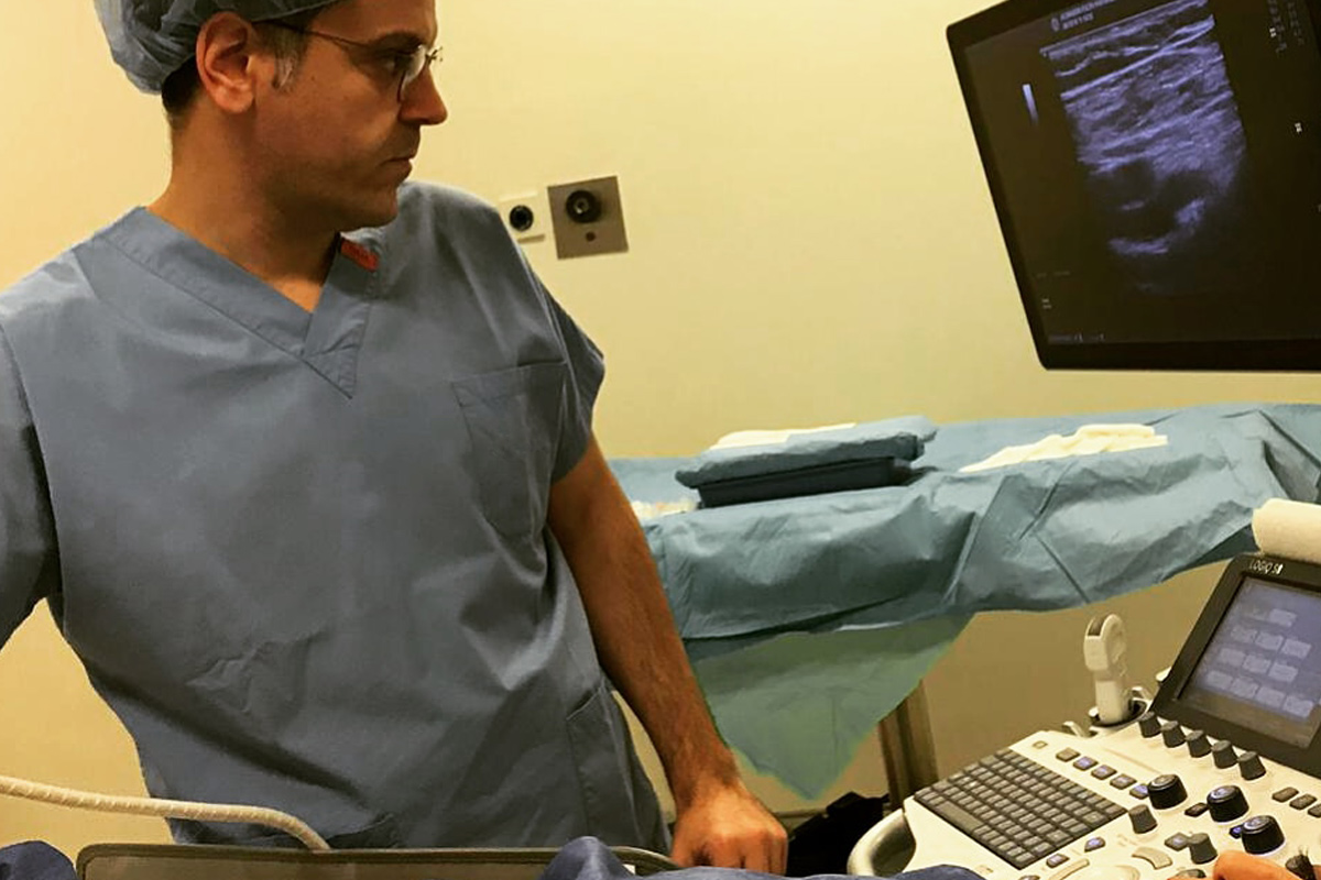
• The most used diagnostic method (it can even be called a scanning method) is venous Doppler ultrasound. The procedure takes about 15-20 minutes, and during that, the patient is not exposed to radiation and is not in a closed environment as in MR -tomography scans. It can also be performed safely in pregnant women.
• Tomography and venography can also be used in advanced and complicated cases.

What is the Purpose of Varicose Vein Treatment?

The treatments used in the treatment of varicose veins are aimed at the symptoms of the disease. The main purpose of treatment is to relieve symptoms such as pain, restlessness, cramps, to eliminate appearance complaints and to prevent recurrence of symptoms through post-treatment follow-up.

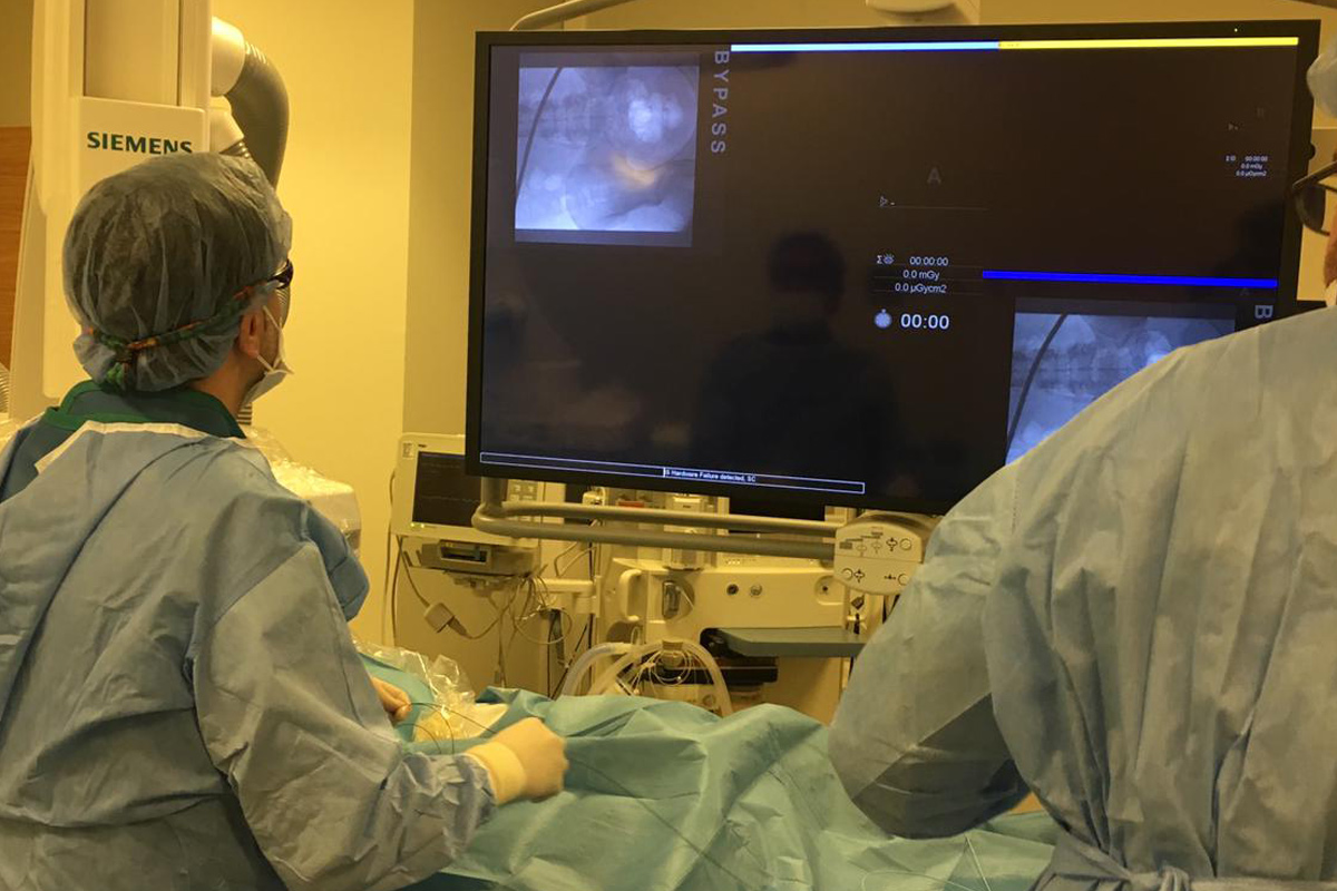
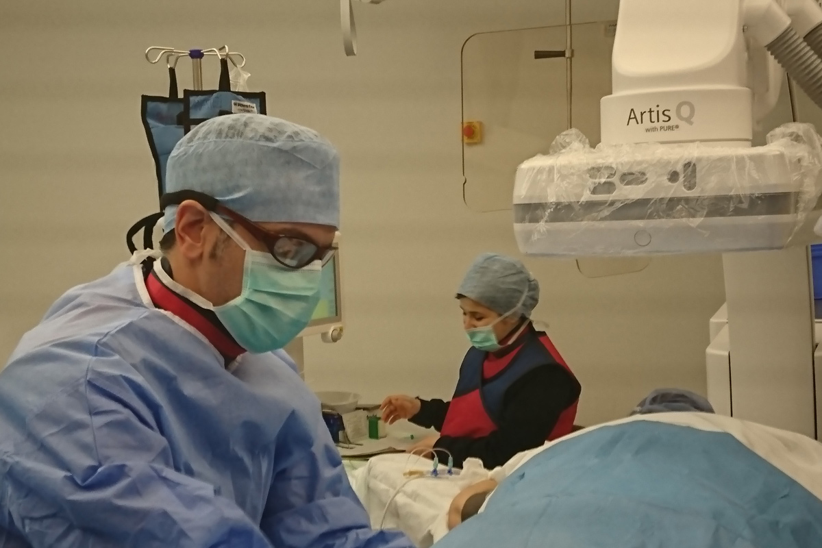
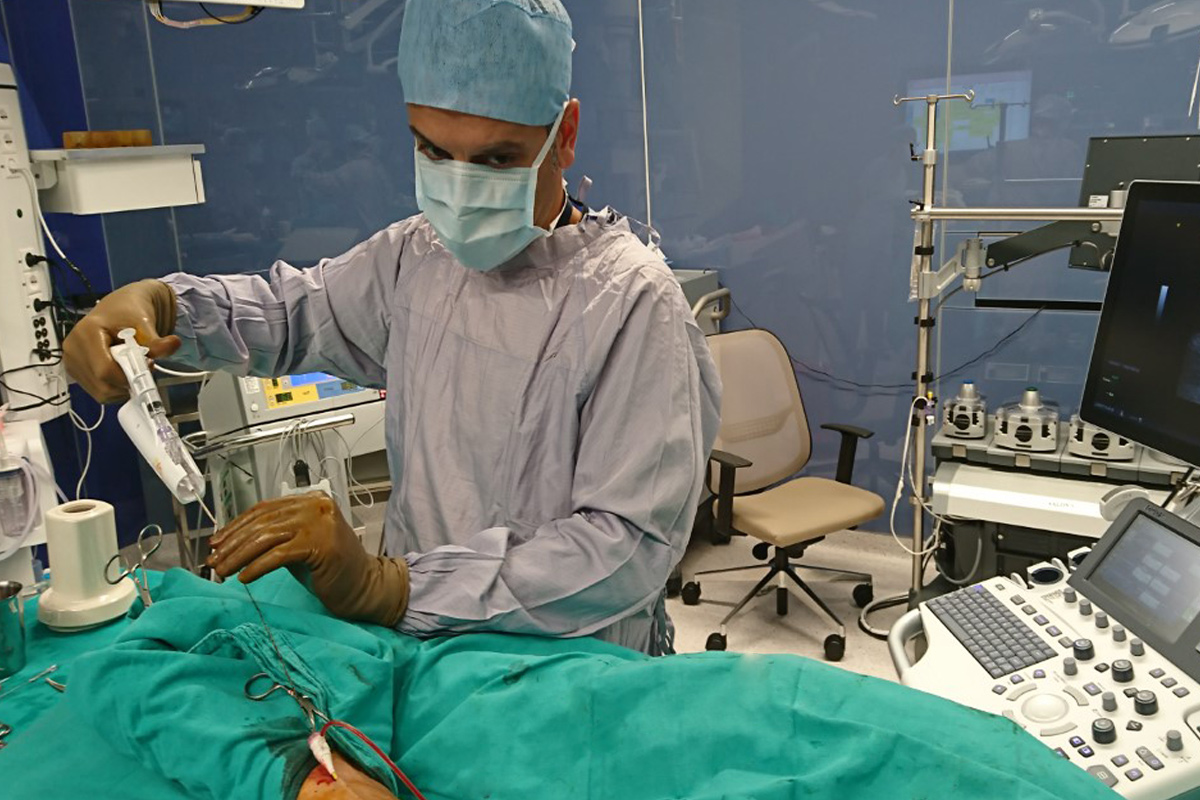
What Methods are Used in the Treatment of Varicose Veins?

We can consider the treatment of varicose veins from two points of view: interventional and medical (medication therapy).

• Interventional Methods
1. Classical Surgery
2. Endovenous Laser Ablation (EVLA)
3. Endovenous Radiofrequency Ablation (RFA)
4. Mechanical Ablation and Foam Sclerotherapy
5. Venaseal method
6. Aesthetic Purposes: Sclerotherapy (foam therapy), laser therapy, radiofrequency therapy

• Medical Therapy
1. Medication Therapy
2. Compression Socks

Varicose Veins and Chronic Venous Insufficiency

Tags: varicose veins and chronic venous insufficiency, varicose veins, what is varicose veins, venous insufficiency, vein, tortuous vein, reflux, valve failure, capillary varicose veins, large varicose veins, genetics, family factors, swelling in the legs, visible veins

Pulmonary Embolism

Pulmonary Embolism

What is Pulmonary Embolism?

Pulmonary embolism (PE) is the term used to describe the clot (thrombus) that has formed in the deep veins of the leg, breaks off from the site where it formed, and occludes the pulmonary artery. Depending on the size of the piece or pieces that have broken off from the clot in the leg, PE can cause an occlusion in the main body of the artery or in one or more of its branches. When one of the main arteries is blocked, it is called a “massive pulmonary embolism” and the situation is very serious.

Who are the candidates for PE?

The source of PE in almost all cases is deep vein thrombosis (DVT). A thrombus (blood clot) that has formed in the deep veins of the leg detaches there, occluding the pulmonary artery and causing PE. People who are at risk for DVT are therefore also at risk for PE.

Apart from these, fatty substances in the marrow of the broken bone, amniotic fluid during pregnancy or birth, and drug abuse are also rare causes that can lead to PE.

What are the symptoms of PE?

Depending on the prevalence and severity of the development of PE, one or more of the following symptoms may occur:

• Breathing difficulties
• Achy chest pain
• Coughing
• Coughing up blood (hemoptysis)
• Palpitations
• Low blood pressure
• Sudden cardiac arrest

How is PE diagnosed?

Doppler ultrasound is initially performed to diagnose DVT in a patient who has possible symptoms and who is suspected of having PE. However, Doppler ultrasound does not reveal PE, it only reveals the presence/absence of DVT, which can cause PE.
Although the D-dimer test rises in PD, it can also rise in many other conditions. Therefore, by itself, it is not conclusive for the diagnosis of PE. However, it may play a role in the clinical course of the patient diagnosed with PE.

Echocardiogram is a very useful, simple, painless, and inexpensive method for diagnosing pulmonary artery embolism. However, the definitive diagnosis of PE is made by a pulmonary angiogram, which is performed using computed tomography.

Pulmonary Embolism

What are the treatment options of PE?

1. Medication Therapy
Anticoagulant (blood thinning) medications: The diagnosed patient should begin taking these medications immediately. They are available in tablet form (Coumadin or new generation anticoagulants) or as injections (heparin or low molecular weight heparin). The effect of the injection forms starts immediately after administration, while the effect of the pill forms starts 12-48 hours after ingestion. The effect of anticoagulant medications used in long-term treatment should be monitored closely because of the risk of re-clotting and bleeding.
Systemic Thrombolytic Therapy: this involves the intravenous administration of various clot-dissolving medications. Its effect is more pronounced than that of anticoagulants, but because the risk of major hemorrhage is higher than with anticoagulants, this is a treatment method that is almost no longer used.

2. Surgical Treatment
In some cases, it is possible to surgically remove the piece of clot that has lodged in the pulmonary artery. However, this operation, which is quite risky, is used only as a last option.

3. Percutaneous Interventions
These are the most widely accepted methods today. In these procedures performed in the angiography room, the patient is placed on the face and an incision is made in the popliteal vein. Depending on the condition/severity of the patient and the disease, one or more of the following procedures may be planned.
• Applications for DVT
• Pulmonary thrombolytic therapy: injection of clot-dissolving drugs into the pulmonary artery.
• Thrombolytic therapy with ultrasound accelerated catheter: This is a method that uses high-frequency ultrasound waves while simultaneously delivering clot-dissolving medication into the pulmonary artery.
• Percutaneous aspiration thrombectomy: It is possible to completely aspirate the clot with a device guided to the pulmonary artery.

Tags: pulmonary embolism, vein, coagulation, pulmonary thrombosis, filter, shortness of breath, chest pain

Deep Vein Thrombosis

Deep Vein Thrombosis

What is DVT?

Thrombosis in ancient Greek means “lump.” Today, thrombosis means the occlusion of a blood vessel by a blood clot (thrombus). Deep vein thrombosis (DVT) is an occlusion in deep veins caused by blood clots.
The incidence of this condition, which most commonly occurs in the veins of the legs, is 1-2 per 1000. This condition, which usually occurs between the ages of 40-50 and later, can also be seen in young people, but is rare.

In which situations does DVT occur?

Long periods of inactivity (e.g., after orthopedic treatments, intensive care processes, or major surgery), long travels, cancer itself or chemotherapy used in cancer treatment, pregnancy and use of contraceptive pill in women, genetic hemorrhagic or hypercoagulability disorders (protein C-S deficiency, such as the factor V Leiden mutation) are among the factors that increase the risk of DVT.

What are the symptoms of DVT?

The occlusion can usually start from the veins in the lower knees and sometimes from the veins in the knees and progress to the main vein in the abdomen (inferior vena cava). The closer the affected vein is to the heart, the more severe the clinical findings. Complaints such as a feeling of tightness and hardness, swelling, pain, warming and discoloration in the legs occur in people with DVT.

How is DVT diagnosed?

The mostly used examination method is Doppler ultrasound. Doppler ultrasound, a method based on the principle of imaging the vessels and their contents using sound waves, is a painless, simple and inexpensive examination method. While it can be used to make a definitive diagnosis in leg veins, other methods may be required for a definitive diagnosis in inguinal and abdominal veins.

In addition to Doppler ultrasound, examinations such as venography, computed tomography or MR are imaging methods used in cases where the diagnosis cannot be made or additional examinations are required.

What are the complications of DVT?

There are two complications of DVT: one that can occur in the early phase, and one that can occur in the late phase.

Pulmonary embolism is when the clot in the vein detaches and is thrown into the pulmonary artery. The symptoms of this condition, which ranks first among preventable hospital deaths, vary depending on the size and prevalence of the clot or clots that cause the occlusion.

The complication that may develop in the late phase is post thrombotic syndrome (PTS). PTS Post thrombotic syndrome is a condition that occurs in 25-75% of patients who have deep vein thrombosis and do not receive appropriate treatment. It presents with symptoms such as leg swelling, pain, and skin thickening, but is more common in high-grade (femoral and abdominal) venous thrombosis. Therefore, to prevent PTS in high-grade DVTs, it is important for long-term success to dissolve the clot with interventional methods in the early stages.

What methods are used in the treatment of DVT?

The conventional treatment for DVT is anticoagulation with heparin or low-molecular-weight heparin, followed by oral anticoagulants. This treatment prevents clot spread and embolization but does not reduce clot mass. In the vast majority of patients treated with the classical method of treatment, post thrombotic syndrome (PTS) develops.

Because thrombolytic therapy dissolves the formed clot in the early phase, the development of PTS can be prevented. Systemic thrombolytic therapy (intravenous administration of blood thinners throughout the body) has been abandoned in many centers because of the potential for bleeding and uncertainties in dosing and administration.

New methods are used with the development of modern medicine. It is possible to administer thrombolytics to the clot with “catheter-directed thrombolytic therapy” or dissolve the clot by medications and catheters with the method of “pharmacomechanical thrombolysis” or directly aspirate the clot with a high rotational force with “aspiration thrombectomy” using mechanical aspiration catheters. Time is very important in these new treatment methods. After the occurrence of DVT, the clot organizes and hardens over time, and then, it is not possible to completely dissolve the clot with the new treatment methods. The ideal period is the first 30 days. After this period, the success of treatment decreases.

Deep Vein Thrombosis

Tags: deep vein thrombosis, coagulation, clot-dissolving treatment, thrombolytic therapy, percutaneous local thrombolytic, clot aspiration, pulmonary embolism, pulmonary thrombosis, filter placement, blood thinners, leg swelling

New Treatment Methods for Deep Vein Thrombosis and Pulmonary Embolism

New Treatment Methods for Deep Vein Thrombosis and Pulmonary Embolism

With the development of modern medicine, percutaneous procedures have been used to treat both deep vein thrombosis (DVT) and pulmonary embolism (PE). Through these treatment methods, the incidence of PE, which may develop in the early phase due to DVT, is reduced and post thrombotic syndrome, the most common problem of these patients in the late phase, can be prevented. Also, the need for intensive care in the early phase is eliminated in patients with PE, reducing the risk of mortality and morbidity and preventing possible chronic lung disease that may develop in the long term.

New Treatment Methods for Deep Vein Thrombosis and Pulmonary Embolism

These treatments, performed under local anesthesia in the angiography room, are completely painless. The patient feels only the local anesthesia used during the first injection. Both ultrasound and angiography are used in these procedures, which are performed face down.

New Treatment Methods for Pulmonary Embolism

By establishing an access to the vein on the knee with ultrasound control, the catheter is advanced with a guide wire after passing the occlusion.

First, a venography is performed and the part of the vein occluded with a clot is traced. Then the clot is dissolved/aspirated and the vein is dilated using the following appropriate methods.

1. AngioJet Pharmacomechanical Thrombectomy
The “Angiojet device,” which is advanced through the clot, allows simultaneous dissolution and aspiration of the thrombus by infusion of a clot-dissolving drug (thrombolytic) and a high-velocity jet of saline.

It can be used for all types of deep vein thrombosis with different catheter diameters and lengths. It can also be used safely and effectively in the treatment of pulmonary embolism.

Because the clot is aspirated while it is dissolved during the procedure, the risk of pulmonary embolism is very low. The risk of major hemorrhage is also very low because the dose of thrombolytics and blood thinners used is very low.

2. Catheter-directed, Ultrasound-assisted Thrombolysis

The ultrasound waves used in this method in which the organized clotting fibers are separated and the effect of the thrombolytic process is enhanced.

In other words, the ultrasound waves facilitate the penetration of the clot-dissolving drug into the clot, thus enhancing its effect. Another substantial aspect of thrombolytic therapy is the dissolution of clots that have remained behind the venous valves.

3. Percutaneous Aspiration Thrombectomy

The treatment “aspiration thrombectomy” is gaining importance for patients who cannot take thrombolytic (clot-dissolving) medications.

Through this catheter with advanced technology, which consists of a spring system that runs over the guide wire and rotates at more than 4000 revolutions per second, the clot in the vein is quickly and actively removed, and patients have the opportunity to be treated without taking anticoagulants.

While almost complete treatment is possible for thrombosis detected in the early phase (in the first 14 days), the success rate of treatment decreases in parallel with waiting and hardening of the clot in the following weeks.

Tags: new treatment methods, interventional treatment, thrombectomy, clot aspiration, thrombolytic therapy, clot lysis treatment, doppler, ultrasound, venography, local anesthesia, stent

Medical Therapy

Medical Therapy

Medical Therapy and Management of the Therapy in Peripheral Vascular Diseases

The treatment of peripheral vascular disease should be performed by teams that are experts in the field and who can apply and know well all the techniques in a wide spectrum, from preventive treatments to surgical options. In Europe and the United States there are doctors who work as “vascular specialists and surgeons”, but in our country there is some confusion in the adoption of the treatment of vascular diseases. However, the most correct way today is for cardiovascular surgeons who have been following and treating vascular disease since residency to provide treatment as a team, along with cardiologists and radiologists.

Elimination of Risk Factors and Lifestyle Changes

While treating diseases of peripheral arteries, it is necessary first to eliminate the causative factors. For this purpose, the following points are important.
• Quitting smoking
• Weight loss
• Regular diet
• Control of diseases such as high blood pressure, hyperlipidemia and diabetes
• Regular exercise

Medical therapy:
In patients who take these precautions but still have pain on walking and at rest, medical therapy is needed. The main medications used in medical therapy are as follows.

• Acetylsalicylic acid (ASA): it is an antithrombotic medication marketed under names such as Aspirin, Coraspin, Ecopirin. It is a blood thinner that works by preventing the cells responsible for blood clotting from sticking together. It is a medicaiton that increases the likelihood of bleeding. It can cause relatively minor bleeding, such as in the mouth, nose, and urinary tract, as well as serious conditions, such as bleeding in the brain. However, because gastrorrhagia can be caused by both blood thinners and contact, it is recommended that it be used in conjunction with gastroprotectants.
• Clopidogrel: It is a stronger blood thinner than ASA. In cases where ASA is not sufficient, it may be recommended to use alone or in combination. As with ASA, Clopidogrel also increases the risk of bleeding.
• Cilastazol: It is a drug sold in the market under the name Pletal. It is a very potent drug that has both an inhibitory effect on blood clotting cells and a vasodilator effect. It is expected to increase walking distance by 500% within 1 month. The most likely side effect is headache. If these headaches, which are usually transient, persist, tolerance is expected to develop by reducing the dose.
• Pentoxifylline: It is a pharmacological agent sold under the name Trental, but it is not as commonly used as it once was.