• 7/24 Hastanemize Ulaşın
  • Hastane Randevu : 0212 919 60 00

Category Archives: Heart Health and Heart Surgery

Diagnostic Procedures

DIAGNOSTIC PROCEDURES IN HEART DISEASES

Exercise Test (Stress Test)

The exercise test can be applied in two ways.

– Exercise ECG (Electrocardiography)
– Stress ECHO (Echocardiogram) (with exertion or medication)

a. Exercise ECG:

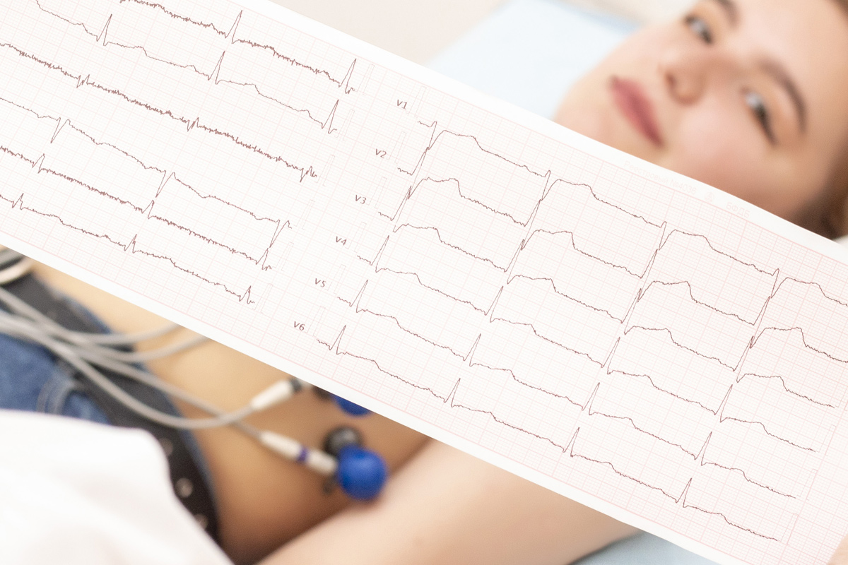
Continuous measurement of electrocardiography of the heart is called “exercise test” while the patient suspected of having stenosis of the cardiac vessels walks at a fast pace on the treadmill.

b. Stress Echocardiogram:

An ultrasound examination (ECHO) of the heart is performed before and immediately after the exercise test in some special cases, which increases the accuracy of the diagnosis of heart disease compared to the normal exercise test.

A 4-hour fasting period is required for the exercise test. It is advantageous for patients to come with athletic shoes and tracksuits if possible.

Echocardiography and Color Doppler

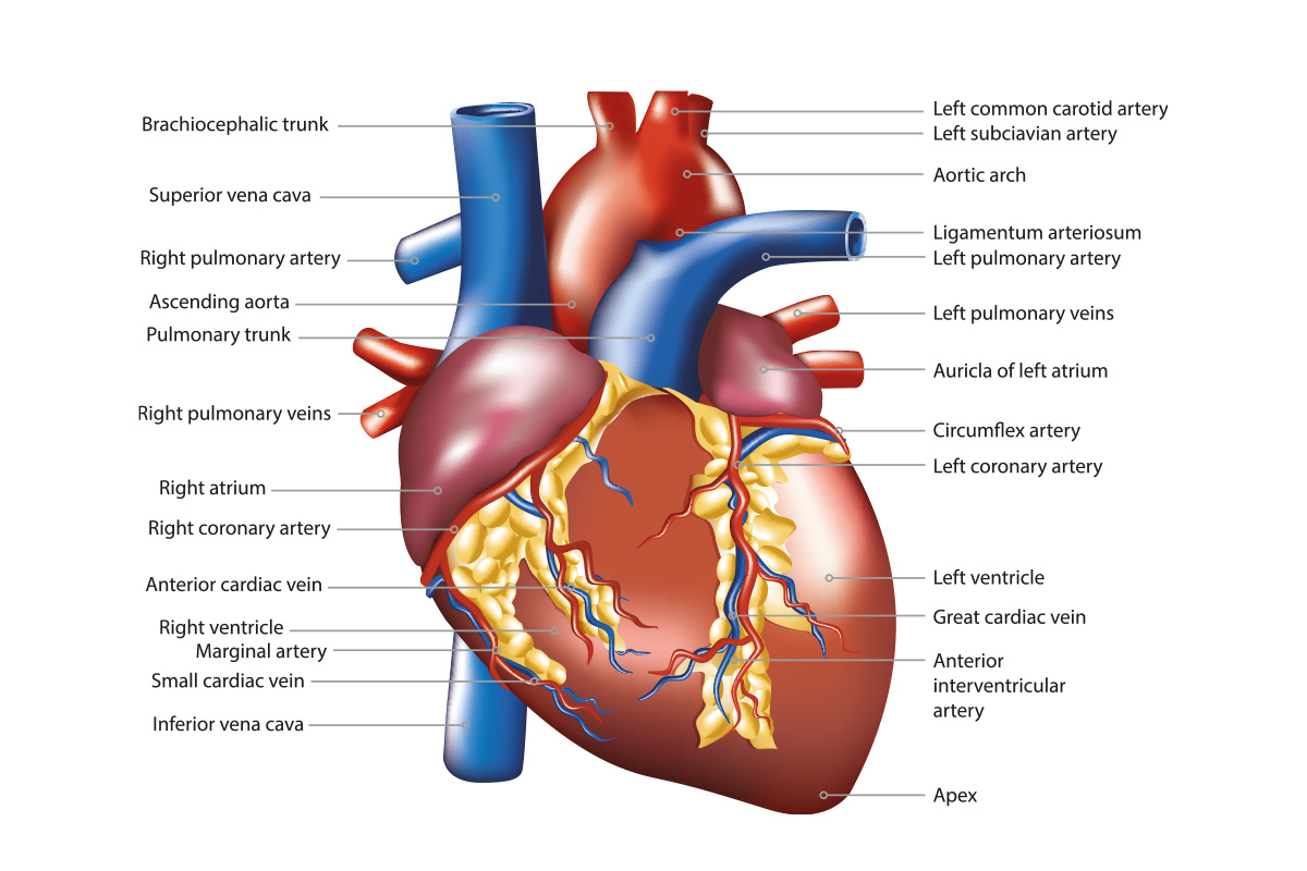
The technique that converts the structure of the human heart, the diameters of its chambers, the working capacity of the myocardium, and, in particular, the structure and functions of the heart valves into live images using the ultrasound wave method is called echocardiography (ECHO). No preparation or fasting is required for this test and the entire test can be performed in a short time.

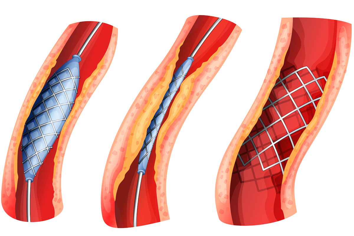
Cardiac Catheterization and Coronary Angiogram

With the cardiac catheterization and coronary angiogram, it is possible to closely observe the structure of the heart, pressures in the heart chambers and large vessels, stenosis, and insufficiencies of the heart valves, and especially stenosis or occlusions of the heart vessels. Thanks to this technology, separate radiological images of each of the heart vessels are taken from different angles. The procedure usually takes between 10 and 20 minutes, and bed rest is required for 4-6 hours after the procedure. It is performed with the help of a catheter that is passed from the femoral artery to the heart under local anesthesia. During cardiac catheterization, the pressures in the heart are measured and at the same time the contractile function of the heart is examined by injecting a dye (contrast substance) into the left heart chamber.

Holter Monitor (Heart Rate or Blood Pressure Holter)

Heart rhythm is monitored and recorded for 24 hours with a device called Holter. This device can be used to diagnose cardiac arrhythmias.
The blood pressure Holter device also records fluctuations in the patient’s blood pressure values for 24 hours, during the day, under real conditions and during sleep.

Electrophysiological Study

It is the most definitive method of diagnosing cardiac arrhythmias. Thanks to this procedure, which is performed with the help of a special catheter passed under local anesthesia from the femoral vein to the heart, the locations of arrhythmias that cause the heart to work too fast (tachycardia) or too slow (bradycardia) are determined by ‘millimetric’ measurements and the ‘map of arrhythmia’ required for definitive treatment is created. This method, which requires very advanced knowledge and technologies, can be applied only in a few centers in Turkey.

Cardiac MRI

It is an imaging technique that can show the structure of the pericardium, heart chambers and valves, myocardium and large arteries coming from the heart, as well as some heart diseases (such as stenosis, vasodilatation, calcinosis) in an extremely simple and harmless way. The processing time is about 15 minutes and requires no prior preparation.

Nuclear Cardiology

a) Myocardial Perfusion Scintigraphy (Thallium Stress Test)

It is a more sensitive method than the exercise test to determine if there is occlusion or stenosis in the vessels supplying the heart. Thallium, a radioactive substance given intravenously, is used to examine the blood supply (or nutrition) to the myocardium is examined at rest and during exercise. A fasting period is required for the test.

b) MUGA: It is the most accurate method of measuring the pumping capacity of the left ventricle. Before the procedure, the red blood cells of the blood are marked with a radioactive substance called technetium. Cardiac function is measured by simultaneous imaging with an ECG scan.

Symptoms of Heart Disease

Symptoms of Heart Disease

1. Chest Pain (Angina)

The sensation of pressure and tightness in the anterior part of the chest, and especially the pain that is triggered by exertion or physical activity and disappears completely at rest, gives the patient and the doctor a clue to heart disease. This pain can sometimes spread or is felt mainly in the left shoulder, the inside of the left arm, the neck, the lower jaw and between the scapulae (shoulder blades). These also usually occur with exertion or physical activity (e.g., walking, running) and are relieved by rest (within a short period of 5 minutes). However, if the same pain occurs while sitting, eating, or sleeping, this is a sign of more serious heart disease (myocardial infarction). This situation requires an immediate ambulance ride to the nearest cardiac center or hospital emergency department. Since it is a life-threatening situation, the most effective intervention can be made in hospitals.

2. Shortness of Breath

It is the sensation of breathing rapidly and yet not being able to breathe easily (dyspnea/air hunger) when walking or running. As with symptoms of chest pain, shortness of breath can also occur at rest and may be a sign of serious heart disease. However, some lung diseases (such as asthma and emphysema) can cause similar symptoms, and it can be very difficult to detect whether the actual cause is from the heart or the lungs. In such cases, it may be necessary for a cardiac surgeon to perform further tests (and sometimes even work with a pulmonologist) to determine the patient’s true problem.

3. Tachycardia

It is a sensation of palpitations felt in the chest, where the heart is located, as a result of a sudden increase in heart rate (e.g., 100-300 beats per minute) or a decrease (e.g., 30-40 beats per minute) or due to irregular heartbeats.

4. Syncope

It is a condition of sudden loss of consciousness, falling to the ground and regaining consciousness after a short time, usually while standing. It may be a sign of very serious heart disease or may be due to many other less serious causes.

5. Swelling in the Feet (Edema)

It is a swelling due to excess fluid in both ankles, the forefoot, or the front of the lower leg. It may be due to advanced heart failure and liver and kidney disease. This must definitely be examined.

Anatomy

General Anatomy

The heart is located behind the anterior chest wall, midway between the two lungs. It is composed of muscular tissue. It is a vital organ whose main function is to pump blood.

Front view of the heart

Front view of the heart; After removal of the anterior parts: the left ventricle, the right ventricle and the right atrium:

It consists of four chambers, an atrium and a ventricle on the right and left sides. The tricuspid valve separates the right atrium and the right ventricle, and the mitral valve separates the left atrium and the left ventricle. The aortic valve is located between the end of the left ventricle and the beginning of the aorta, the largest artery of the human heart. Similarly, the pulmonary valve is located between the right ventricle and the pulmonary artery. The veins (inferior vena cava and superior vena cava), which bring blood from the whole body, are connected to the right system of the heart. This blood leaves the right system through the pulmonary artery. Blood returning from the lungs through the pulmonary veins circulates to the left atrium and left ventricle and is pumped throughout the body through the aorta.

The outer surface of the heart is surrounded by a membrane called the pericardium. Between this membrane and the heart is a very small amount of lubricating fluid to allow the heart to move freely during its function.

How to Prevent Cardiovascular Diseases?

How to Prevent Cardiovascular Diseases

Dealing with risk factors is very important. It is not possible to change risk factors such as age, gender, and family history. However, other factors can be addressed. Your doctor will guide you in this regard. You should stop smoking, control high blood pressure, keep diabetes within normal limits, exercise, diet if you have high cholesterol, and if your doctor recommends medication, take it.

How is the diagnosis made?

The most important thing in coronary artery disease is to treat the narrowing of the coronary arteries before it causes an infarction. This is accomplished primarily by observing the condition of the coronary arteries. Although there are various methods used for this purpose (stress test, myocardial scintigraphy, etc.), the coronary angiogram is still the best diagnostic method and is considered the gold standard in patients with suspected vascular disease. A coronary angiogram involves visualizing the coronary arteries and taking images.

Treatment of Cardiovascular Diseases

Treatment of Cardiovascular Diseases

If significant narrowing and/or occlusion of the coronary arteries is detected, balloon angioplasty can be performed in the same session or later if necessary. Balloon angioplasty involves inflating a specially made balloon for a short period of time and widening the narrowing of the vessel. The balloon can be used for more than one stenosis in the same vein or for stenoses in more than one vein in the same session or in different sessions. If necessary, a stent is placed in the area using a balloon in addition to it.

In the balloon procedure, a catheter with an inflatable balloon at its tip is inserted into the area of stenosis (A). After it is placed in the narrowed area, the balloon is inflated and the stenosis is opened (B).

In cases not suitable for balloon angioplasty, bypass surgery or medication therapy may be recommended.

In bypass surgery, a vessel that serves as a bridge is inserted between the front and back of the site where the stenosis begins. This vessel can be the patient’s own saphenous vein or an artery (lima) from the chest. This allows blood to flow beyond the narrowed or occluded area with the help of this bridge.

Here we see LIMA bypass to the left anterior descending artery (LAD) and saphenous vein bypass to the right coronary artery (RCA).

Medications used for coronary artery disease dilate the coronary arteries, reduce the load on the heart, prevent the formation of blood clots in this area, or lower cholesterol levels, which have a very negative effect on atherosclerosis. The medicines should be taken continuously and under the supervision of a doctor.

Here we see that the leg vein (saphenous vein graft) is anastomosed to the left anterior descending artery (LAD). First, one end of the graft is sutured to the LAD, then the other end is sutured to the aorta. Thus, blood coming from the aorta bypasses the occluded area on the other side of the artery and supplies the heart.

Unfortunately, neither the medications used in the treatment of coronary artery disease nor the balloon and bypass can eliminate the vascular disease. Therefore, new stenoses may occur in the same region or in different regions of the coronary vessel, or a mild stenosis may progress and become a severe stenosis and cause problems. For this reason, it is very important for patients to take their medications regularly and to come in for checkups at times recommended by their doctor to eliminate risk factors and whenever they have other symptoms to detect problems early.