• 7/24 Hastanemize Ulaşın
  • Hastane Randevu : 0212 919 60 00

Category Archives: Peripheral Vascular Diseases

Medical Therapy

Medical Therapy

Medical Therapy and Management of the Therapy in Peripheral Vascular Diseases

The treatment of peripheral vascular disease should be performed by teams that are experts in the field and who can apply and know well all the techniques in a wide spectrum, from preventive treatments to surgical options. In Europe and the United States there are doctors who work as “vascular specialists and surgeons”, but in our country there is some confusion in the adoption of the treatment of vascular diseases. However, the most correct way today is for cardiovascular surgeons who have been following and treating vascular disease since residency to provide treatment as a team, along with cardiologists and radiologists.

Elimination of Risk Factors and Lifestyle Changes

While treating diseases of peripheral arteries, it is necessary first to eliminate the causative factors. For this purpose, the following points are important.
• Quitting smoking
• Weight loss
• Regular diet
• Control of diseases such as high blood pressure, hyperlipidemia and diabetes
• Regular exercise

Medical therapy:
In patients who take these precautions but still have pain on walking and at rest, medical therapy is needed. The main medications used in medical therapy are as follows.

• Acetylsalicylic acid (ASA): it is an antithrombotic medication marketed under names such as Aspirin, Coraspin, Ecopirin. It is a blood thinner that works by preventing the cells responsible for blood clotting from sticking together. It is a medicaiton that increases the likelihood of bleeding. It can cause relatively minor bleeding, such as in the mouth, nose, and urinary tract, as well as serious conditions, such as bleeding in the brain. However, because gastrorrhagia can be caused by both blood thinners and contact, it is recommended that it be used in conjunction with gastroprotectants.
• Clopidogrel: It is a stronger blood thinner than ASA. In cases where ASA is not sufficient, it may be recommended to use alone or in combination. As with ASA, Clopidogrel also increases the risk of bleeding.
• Cilastazol: It is a drug sold in the market under the name Pletal. It is a very potent drug that has both an inhibitory effect on blood clotting cells and a vasodilator effect. It is expected to increase walking distance by 500% within 1 month. The most likely side effect is headache. If these headaches, which are usually transient, persist, tolerance is expected to develop by reducing the dose.
• Pentoxifylline: It is a pharmacological agent sold under the name Trental, but it is not as commonly used as it once was.

Carotid Artery Disease

Carotid Artery Disease

What is carotid artery disease?

Carotid artery disease is a disease that results from narrowing or occlusion of the carotid arteries that enter the skull from both sides of the neck and supply the brain. It is a very serious disease. It ranks first worldwide in deaths from neurological causes and second in deaths from cardiovascular diseases.

What are the causes of the disease?

Along with family history and genetic factors, smoking, irregular diet, hypertension, hyperlipidemia, and diabetes are the most important causes.

What are the symptoms?

In the initial phase, it causes symptoms such as dizziness, blackout, and amnesia. Subsequently, there may be temporary loss of vision, speech disorders, fainting and temporary loss of consciousness. If paralysis has developed due to the carotid artery disease, movement and sensation disorders and speech disorders may occur in the half of the body on the opposite side of the affected carotid artery.

What is a stroke?

When the blood supply to a part of the brain is interrupted for more than a few minutes, the brain cells in that region are destroyed and the patient develops a loss of vision, speech, movement or sensation in certain parts of the body. If the underlying condition is only temporary, the symptoms it causes may also be temporary, which is called a “transient ischemic attack.” If the lost blood flow does not return and the stroke lasts longer than 24 hours, this situation is called a “stroke.”

What are the diagnostic methods?

Although the initial imaging modality for carotid artery disease is Doppler ultrasound, magnetic resonance (MR) or computed tomography (CT) angiogram studies should also be performed for a definitive diagnosis. Yet, a definitive diagnosis of carotid artery disease can be made with conventional angiography.

Is it possible to treat?

Carotid artery disease can be treated. The method of treatment depends on the degree of stenosis, the symptoms present, and whether a stroke has occurred.

Medical treatment: Anticoagulant and vasodilator treatments can be used in patients with carotid artery stenosis of less than 70% who have not yet suffered a stroke or ischemic attack.

Interventional methods and surgical treatment: it should be used in patients who do not have complete occlusion of the carotid arteries but have stenosis greater than 70%. Angiography involves insertion of a stent into the carotid artery. However, scientific studies show that the rate of long-term dilation of the vessel is higher with surgical treatment and the likelihood of recurrence of the disease is lower. During surgical treatment, the narrowed section of the vessel is opened and cleaned, repaired, and resealed under local or general anesthesia.

To determine which treatment method is appropriate for your condition, it is best to consult your doctor, evaluate the possible side effects, complications and benefit-harm ratio, and decide on the treatment method.

Vascular Emergencies

Tags: carotid artery disease, carotid artery, stroke, paralysis, endarterectomy, stent

Interventional Treatment

Interventional Treatment in Peripheral Vascular Diseases

Management of the Treatment Process

The treatment of peripheral vascular disease should be performed by teams that are experts in the field and who can apply and know well all the techniques in a wide spectrum, from preventive treatments to surgical options. In Europe and the United States there are doctors who work as “vascular specialists and surgeons”, but in our country there is some confusion in the adoption of the treatment of vascular diseases. However, the most correct way today is for cardiovascular surgeons who have been following and treating vascular disease since residency to provide treatment as a team, along with cardiologists and radiologists.

Interventional Methods (Endovascular Procedures)

Interventional Treatment

This is the first-level procedure that should be used in patients with non-healing wounds on the feet and pain at rest, as well as in patients whose legs are to be amputated but for whom attempts are being made to amputate them at a lower level. The choice between interventional methods and surgery is made by evaluating the location and length of the stenosis, the vascular structure, and the general condition of the patient.

Endovascular Methods

All interventional methods are performed in the angiography room under local anesthesia and without incision. The hospital stay and recovery period are shorter than for surgical treatment. However, there is a recurrence risk of stenosis or occlusion in the treated vessels in the early or late phases.

Depending on the location, length, and duration of the occlusion or stenosis, one or more of the following procedures may be used in combination.

• Atherectomy: It is a treatment method based on the principle of aspirating the plaques (calcinosis) formed in the vessels by breaking them up with the help of special catheters. This method is used mainly in cases of vascular occlusion/vascular stenosis below the knee and long vasoconstrictions in the thigh area, as well as in cases of complete occlusion. It may need to be combined with a balloon and/or a stent.
• Balloon angioplasty: this method is effectively used in the treatment of vascular stenosis. It is based on the adhesion to the vessel wall of plaques that cause narrowing of the vessel. It can be performed with medicated or non-medicated balloons. In addition to their mechanical effect, medicated balloons are more effective than non-medicated ones due to the anti-inflammatory effect of the pharmacological agents they create on the vessel wall.

Balloon Angioplasty

• Stent implantation: this method is used mainly in the arteries above the groin and occasionally in the leg arteries. It is not recommended to be placed in vessels below the knee unless absolutely necessary. There are stents made entirely of metal and stent grafts covered with various materials that are compatible with the body.

Interventional Treatment in Peripheral Artery Diseases
Although it is known before the interventional procedure which of the above methods will be used, the final decision as to which method is necessary and will be used is made during the procedure.

Tags: interventional treatment, angiography, catheter, balloon, stent, atherectomy, thrombolytic therapy, angioplasty, endovascular, percutaneous intervention, vascular occlusion, atherosclerosis

Arteriosclerosis

Arteriosclerosis

What is peripheral vascular disease?

Peripheral vascular disease (PVD) is the degeneration of the arterial system in our body due to atherosclerosis. In this degeneration, the interior of the vessel first narrows due to calcinosis and the accumulation of fatty layers on the walls of the vessels, and then with the progression of the disease, complete occlusion occurs.

What are the causes of PVD and in whom does it occur more frequently?

PVD is a disease that usually occurs in advanced age (over 50 years). It occurs more frequently in men than in women. In addition to family history and genetic factors, lifestyle habits such as smoking, lack of exercise, irregular diet, and chronic diseases such as hypertension, hyperlipidemia, diabetes, and kidney failure are factors that increase the incidence.

In which vessels is it most common?

PVD most commonly occurs in the leg vessels of the body. However, the carotid arteries (carotid artery disease), brachial arteries, and visceral organ veins (kidney, intestine, etc.) may also be affected.

What are the symptoms of PVD?

When PVD affects the vessels that supply the organ, there are findings about that organ. It usually occurs in the arteries of the leg and the symptoms vary depending on the degree of narrowing and the location of the occlusions in the leg arteries.

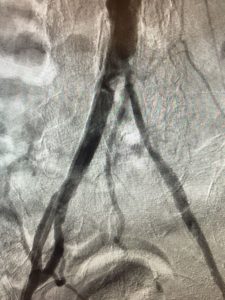
Common complaints can be listed as follows:
• Pain: The pain begins when walking and disappears when resting. It is a cramp-like pain that occurs in the muscles of the affected region due to the undersupply that results from not being able to get enough oxygenated blood to the narrowed area. While the pain of problems in arteries in the upper part of the leg is felt throughout the leg, the problem in vascular occlusion in the knee area is in the region below the knee.
The more severe the occlusion and stenosis, the less exertion the pain causes and the later it subsides. In the advanced stages of the disease, pain at rest without exertion is observed.

Arteriosclerosis
• Wound: depending on advanced undersupply to the legs and feet, wounds may open spontaneously, or a wound opened by a cause such as trauma or a cut may not heal.
• Gangrene: This is the loss of vitality of the limbs when they are undersupplied. Amputation of the gangrenous part is often unavoidable.

Arteriosclerosis

How is PVD diagnosed?

The first step of diagnosis is the examination and the arm and foot pressure ratios. After that, it is necessary to use imaging techniques.

Doppler ultrasound examination of the arteries does not provide precise information about the stage of the disease, but it does provide approximate information about the stage of the disease and the vessels involved.

Angiographic diagnostic techniques must be used to make a definitive diagnosis and guide treatment. Magnetic resonance (MR) or computed tomography (CT) angiography can objectively determine the location and degree of stenosis and identify 3D anatomic features.

With conventional angiography, both diagnoses can be made and balloon, stent, or other procedures in cases requiring treatment can be performed during one procedure.

Arteriosclerosis

What are the treatment options?

Since the effects differ according to the stage of the disease, the treatment also differs according to the person. Therefore, the treatment of people with PVD should be planned taking into account the patient’s disease, symptoms, limb vitality, and the person’s social expectations.

In cases of pain at rest, gangrene or a non-healing wound in the foot or leg, interventional treatment that increases blood supply is absolutely necessary because of the risk of losing the limb and perhaps life. However, for patients who have no symptoms other than pain when walking, the treatment method should be chosen with consideration of factors such as the patient’s age, social circumstances, and the extent to which his or her life is affected by the disease. For example, for a 50-year-old, active, working patient, surgical treatment may be recommended to treat pain on walking 1 km, whereas for an 80-year-old patient who leaves home 1-2 times a week and has pain on walking 250 meters, medical treatment and follow up may be recommended.

What should a patient with PVD pay attention to?

It is necessary to eliminate all factors that cause the development of the disease. Family factors are inevitable and unchangeable. However, these patients must stop smoking, exercise and lose weight.

People with PVD are recommended to walk at least 3 times a week for 45-60 minutes at an athletic pace. In addition, factors such as hypertension, hyperlipidemia, and diabetes, which play a direct role in the development of PVD, should be adequately controlled.

Even taking these precautions alone will increase the walking distance of patients by 1.5-2 times within 3-6 months.

Arteriosclerosis

Is medication therapy effective in PVD?

There are drug treatments for PVD that have been shown to be effective. In particular, some blood-thinning and procirculatory medications can increase walking distance by up to 2 times within 3-6 months of their use and relieve existing pain at rest.

The most commonly used medication therapy is the anticoagulant medication aspirin and the medication Pletal (cilastazol), which has vasodilator and blood thinning effects.

In PVD, the medications must be used under the supervision of a physician.

What other treatment options are available for PVD?

Interventional treatment methods and surgical treatment methods are used for patients who do not achieve the expected results despite changes in lifestyle and medication therapy.

Interventional treatment methods are procedures that are accompanied by angiography. It is an effective treatment method widely used in the world and in our country.

Treatment is possible with atherectomy (removal of plaques causing vascular occlusion), balloon and stent systems, which are inserted into the area over a fine wire system during the procedure, depending on the location and length of the occlusion or the stenosis. Patients are discharged 6-8 hours after the procedure or the next day, depending on the length and techniques used during the procedure.

Surgical treatment methods are used in cases with very long or complete occlusion where interventional methods cannot be used or in cases of recurrent stenosis after interventional methods.

There are basically two types of surgical techniques: Bypass and plaque removal. In bypass surgery, a new bridge is created before and after the occlusion that causes peripheral vascular disease, either through veins taken from the body or artificial vessels. In this method, blood is directed beyond the occlusion via an alternative new pathway. Plaque removal involves opening the artery in the area where the blockage is located, cleaning the inside, and then repairing the artery.

Arteriosclerosis

Tags: arteriosclerosis, vascular occlusion, Buerger’s disease, balloon, stent, angiography, bypass, artificial vessel, acute vascular occlusion, endovascular, percutaneous intervention, catheter, Reynaud’s syndrome, Reynaud’s disease, gangrene, leg wound

Vascular Aneurysm

Vascular Aneurysm

What is aneurysm and where can it be seen?

An aneurysm is defined as an artery in the body that bulges to twice its normal diameter. It most commonly occurs in the abdominal aorta, while the popliteal artery is the most common site for peripheral aneurysm.

What are the findings in abdominal aortic aneurysms?

Abdominal aortic aneurysms are generally silent. Mild abdominal pain may occur in half of patients. However, abdominal pain is a very indefinite symptom and can occur with many conditions. Sometimes a pulsatile swelling is felt in the abdomen or groin. Most of these patients are diagnosed during examinations such as ultrasound and computed tomography, which are performed for other reasons.

How is it diagnosed?

Abdominal aortic aneurysm can be diagnosed by ultrasound, magnetic resonance, and computed tomography angiography. Yet, CT angiography is the most useful method to select the treatment method and determine the details of the anatomy of the disease.

Why is abdominal aortic aneurysm important?

Under normal conditions, abdominal aortas 2-3 cm in diameter pose no risk until they reach 4.5-5 cm. However, abdominal aortas larger than 5 cm increase the risk of spontaneous rupture by 50%. Half of the patients with spontaneous rupture die before arriving the hospital, and the risk of dying during the procedure/surgery is about 50% for the half that can arrive the hospital. Therefore, diagnosis and follow-up of abdominal aortic aneurysms and choosing the right treatment at the right time are important.

When is surgical or interventional treatment performed for abdominal aortic aneurysms?

Aneurysms less than 5 cm in diameter should be followed up at 6-month or 1-year intervals. Surgical or endovascular intervention is required in patients with a diameter greater than 5.5 cm, with an increase in diameter of more than 0.5 cm within 6 months, or in patients with a diameter of 4.5-5.5 cm whose pain complaints persist despite all types of medical treatment.

How a surgery is endovascular aneurysm repair (EVAR)?

EVAR is performed in the angiography room and applied only through 5 cm incisions in the groin. This procedure, which can be used in patients with an abdominal aorta with suitable anatomical features, has a lower risk of mortality and fewer complications compared to surgical treatment.

With EVAR, a procedure that can be performed under general or local anesthesia, the procedure time is shorter, and discharge, return to normal life, and recovery are significantly faster than with surgical treatment.

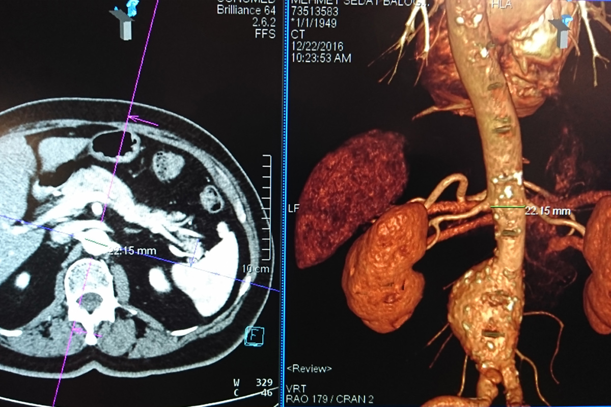
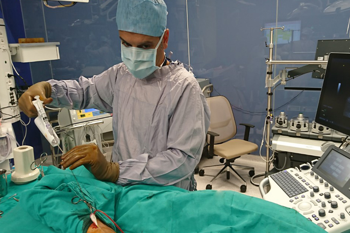
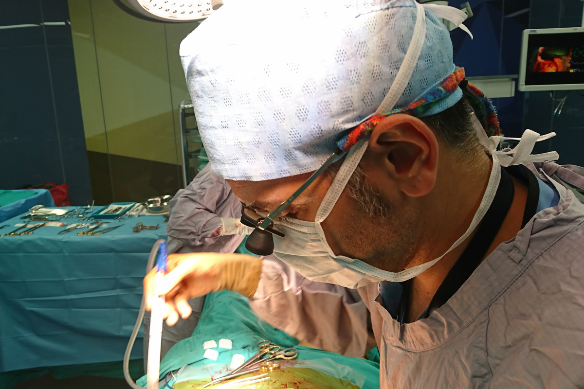
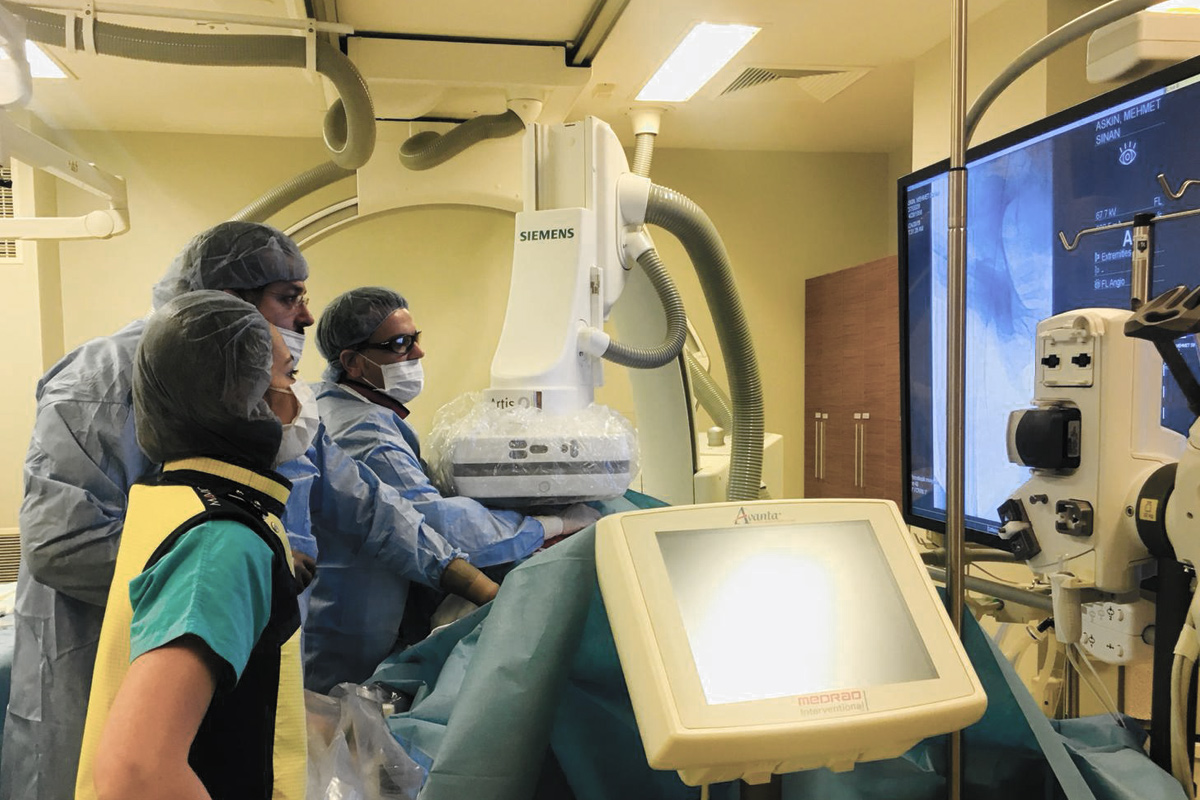
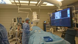
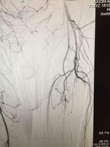
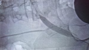
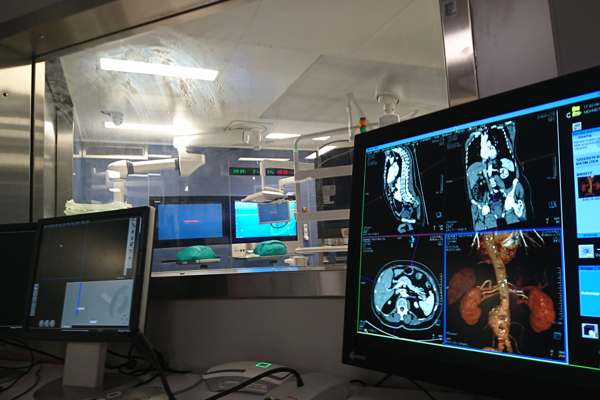
After assessing the anatomical features of the abdominal aorta in 3D with computed tomography, individual and special stent grafts are harvested for each patient. First, both arteries in the groin (femoral arteries) are surgically dissected in the angiography room and the stent grafts harvested specifically for the patient are inserted into the artery in such a way that the vascular aneurysm areas are closed by the procedure through these arteries. Then the incisions are closed by repairing the arteries and completing the process.

Is surgical treatment still performed for abdominal aortic aneurysm?

Although EVAR plays a very important role in the treatment of abdominal aortic aneurysms thanks to technological and technical developments, the need for surgical treatment remains in certain groups of patients.

Although the length of hospital stay, likelihood of death and complications, hospitalization after the procedure, and duration to return to normal life are higher, surgical treatment remains the only option in patients whose anatomical characteristics are not suitable for EVAR.

Aneurysms
Tags: vascular aneurysm, aneurysm, aortic aneurysm, aorta replacement, EVAR, stent, graft, bulging